Cerebral CT scans in trauma.
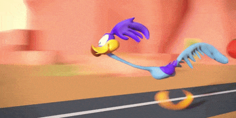
What best describes this cerebral CT scan taken following the head injury of a child?
This is acute (white appearance of acute blood), an extradural (lens shaped because extension restricted by cerebral sutures) and left sided (left side of patient right side of scan).
This patient is on Apixaban and presents following a fall with right sided weakness. What best describes this scan?
Without the restriction of the bony sutures a bleed in the subdural space can extend much further and has a characteristic cresenteric shape.Effacement is just a trendy way to say squished and epidural is the same as extradural.
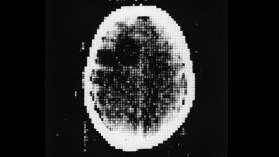
This video is of the cerebral CT of a patient who had a high speed MVA. What does it show?
Rapid deceleration causes widespread shearing forces which give you multiple small bleeds. Despite not seeing much on the CT the patient often has a significant cerebral insult. I thought I made up airbag syndrome--but no--it is where a person doesn't wear a seat belt because they trust their airbag in a crash.
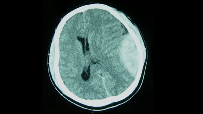
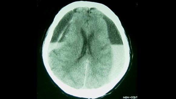
This cerebral CT is of a man who has deteriorated following a fall 10 days ago. What does it show?
It is a large left subdural and is close to isodense. So it is a subacute subdural---the injury occurring about 1-3 weeks before. The mass effect has effaced the left lateral ventricle and squished out all the CSF in the sulci. As the blood in the subdural breaks down it will eventually become hypodense.
Sir Godfrey Hounsfield developed the first Cerebral CT scan. Which of the following statements is untrue?
The teachers got it wrong. Australia has 70 CT scanners per 1 million population compared to the USA with 45 per million. CT scanners first arrived in Australia in 1975.
This is the CT of a child with suspected NAI. What does it show?

This infant had been shaken and has an acute right subdural with associated significant oedema (loss of grey white differentiation). That dark crescent shaped area on the left is the chronic subdural. It does look a bit like an angry face.
This patient was on Clopidogrel and was involved in an MVA. What does it show?

Trauma caused a fair bit of damage but this cut does not show any contusion and that appearance of a potential chronic subdural is an artifact. Probably a bit unfair--but--only a bit.
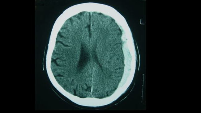
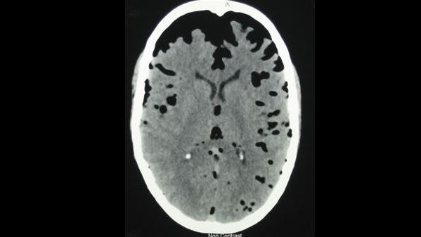
An elderly patient presents with multiple falls and a decrease in LOC. What does this show?
There are subdural haematomas on both sides. Lying supine has allowed the blood to layer out, with the heavier acute blood dropping down due to gravity. The poor old brain is getting squished from both sides.
This man was involved in an MVA and has facial injuries and a headache. What does this CT cut show?
Somewhat surprisingly he had a normal GCS. Air has tracked from fractured frontal and maxillary sinuses into the subarachnoid spaces.
Drilling a hole in the head (trepanation) is the oldest neurosurgical procedure. Which is the following is incorrect?

It is indeed a tulip---the Dutch word for tulip is evidently similar to stupid. Unfortunately the Bosch company was not inspired but the painting. Pity.