Some ECGs you really should know.
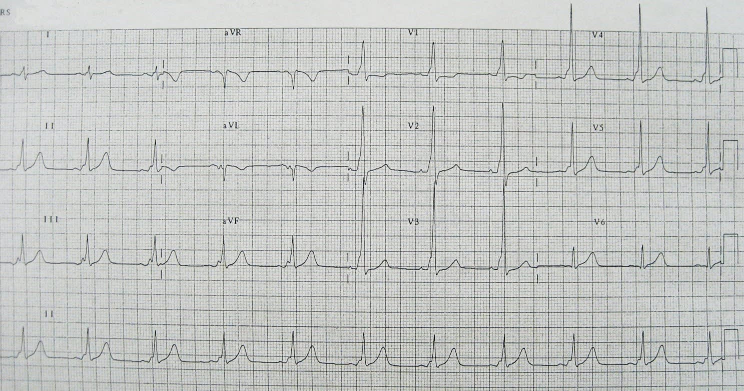
A 62yr old Director of Medical Services presents with central chest pain radiating to his neck. What does the ECG show?
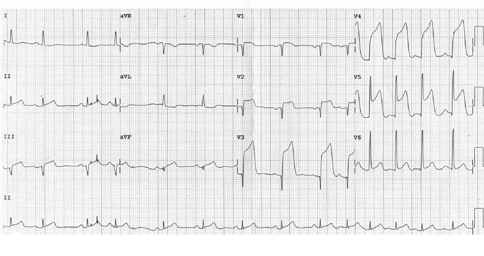
Anterior STEMI.
Anterolateral STEMI.
Severe pericarditis.
Inferoposterior AMI
Pretty straight forward start. ST elevation on almost all chest leads. Needs to have a cardiac catheter to open their artery ASAP. Likely LAD (Left anterior descending) lesion.
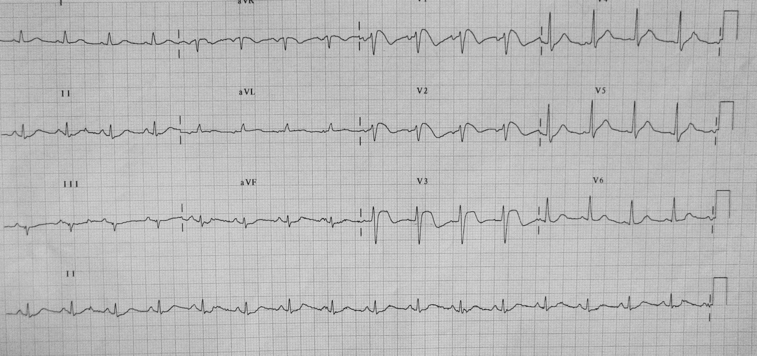
A 28yr old man presents with sudden onset left sided chest pain and shortness of breath. What does the ECG show?

Sinus tachycardia. Non STEMI inferiorly.
Sinus tachycardia. Anterior non STEMI.
Pericarditis.
Sinus tachycardia. s1 q3 t3
S1Q3T3 is indicative of acute cor pulmonate of any cause. First described in 1935. In this clinical presentation it would mean a massive or sub massive pulmonary embolism.
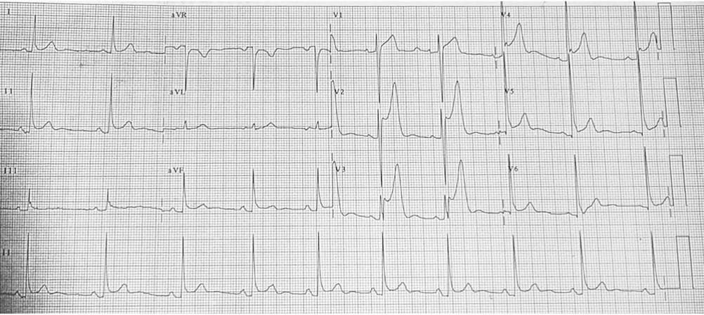
This unusual looking ECG was taken of a 72yr old man who presented with light headedness. What does it show?
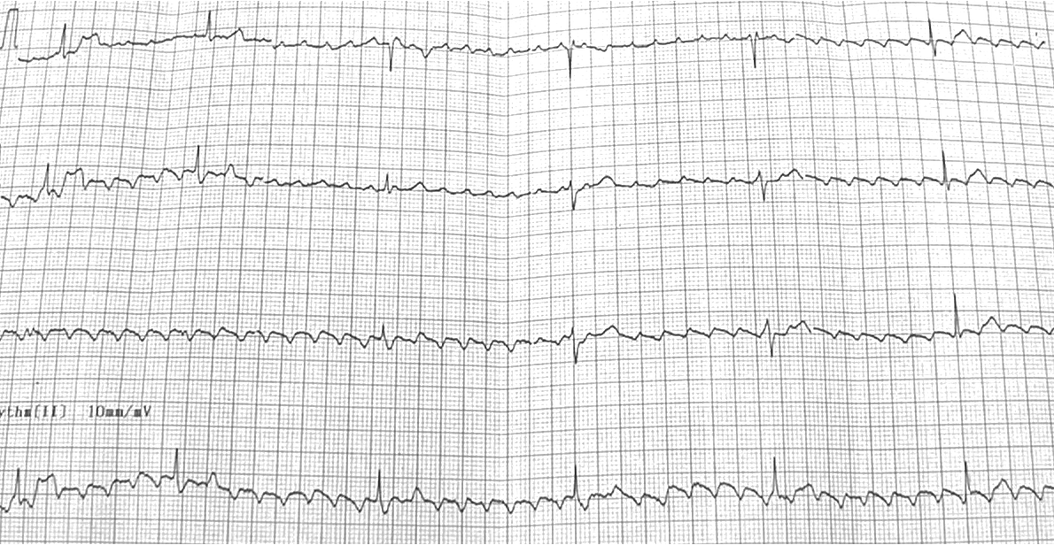
Sinus bradycardia with electrical interference (60Hz)
Complete heart block.
Atrial fibrillation.
Atrial flutter with about 8.1 block.
Flutter waves are often seen best in V1 however here you can see them easily on all leads. At 8.1 block the patient has a heart rate of only about 30 to 40/min.
A previously healthy 30yr old man present with atypical chest pain. The following ECG was taken and repeated 15min later with no change. The initial troponin was normal. What does it show?
Acute pericarditis.
Benign early depolarisation.
Anterior STEMI.
Brugada syndrome.
This is a normal variant seen in healthy young patients. There is often notching of the J point----the "fish hook" appearance that you can see in V4. The ST elevation tends to be more prominent at slower heart rates and disappear in the presence of tachycardia.
What is this man doing?
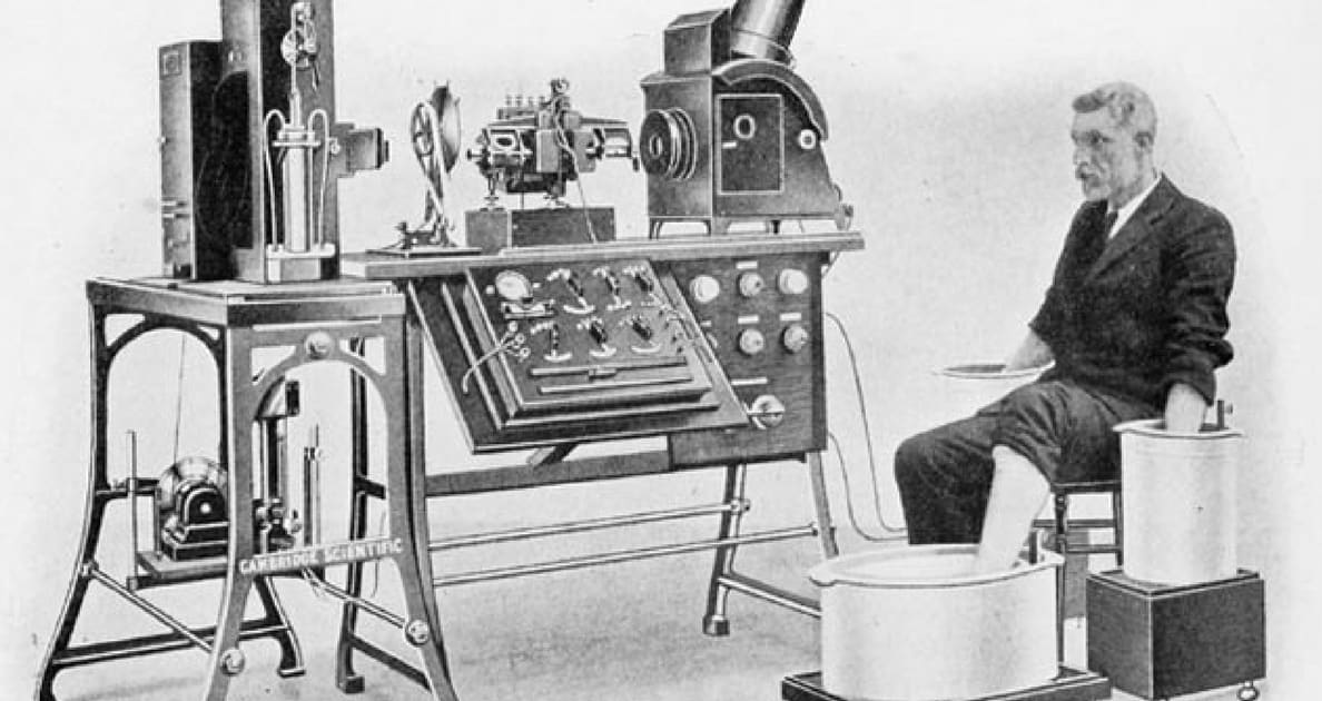
Demonstrating that water and electricity don't mix.
Demonstrating the first machine for cardioversion.
Demonstrating the first ECG machine.
Posing for the first documented photograph of medical research.
It is Willem Einthoven who developed the first ECG machine in 1895. He got the Nobel prize for it in 1924. He looks nervous, as you would with live electricity and your limbs in buckets of water.
A 45yr old man is having an elective operation and had this pre op ECG. What does it show?
Bundle of Cant syndrome.
Vukosovic Ittimani syndrome.
Wolff Parkinson White syndrome.
Lesch Nyhan syndrome.
Note the short PR and slurred upstroke on the QRS (Delta wave). Conduction bypasses the AV node and goes does the Bundle of Kent. Prone to re entrant tachyarrhythmias. Most dangerous is rapid AF as the conduction is much faster down the Bundle of Kent than the AV node.
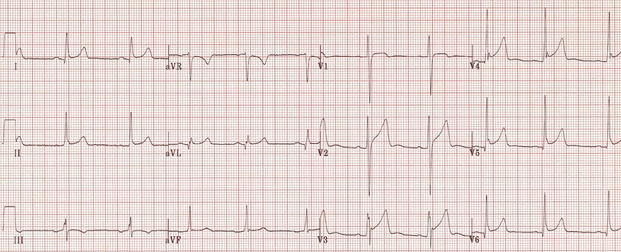
A 56yr old lawyer has recent chest pain which has now resolved What does the ECG show?
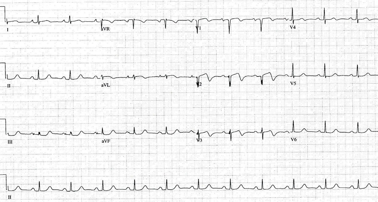
Pericarditis
Wellen's syndrome.
Inferior ischemia.
Normal ECG.
2 Types of Wellens syndrome are described. Type B has dagger like t wave inversion in the chest leads. Type A --like this one---shows "coved shaped" ST elevation in V2 V3. It is seen usually in patients with recent chest pain and these patients have a high risk of developing an anterior AMI. This man had an LAD lesion.
A 16yr old girl with anorexia nervosa and bulemia is getting an ECG when she collapses. What does it show?
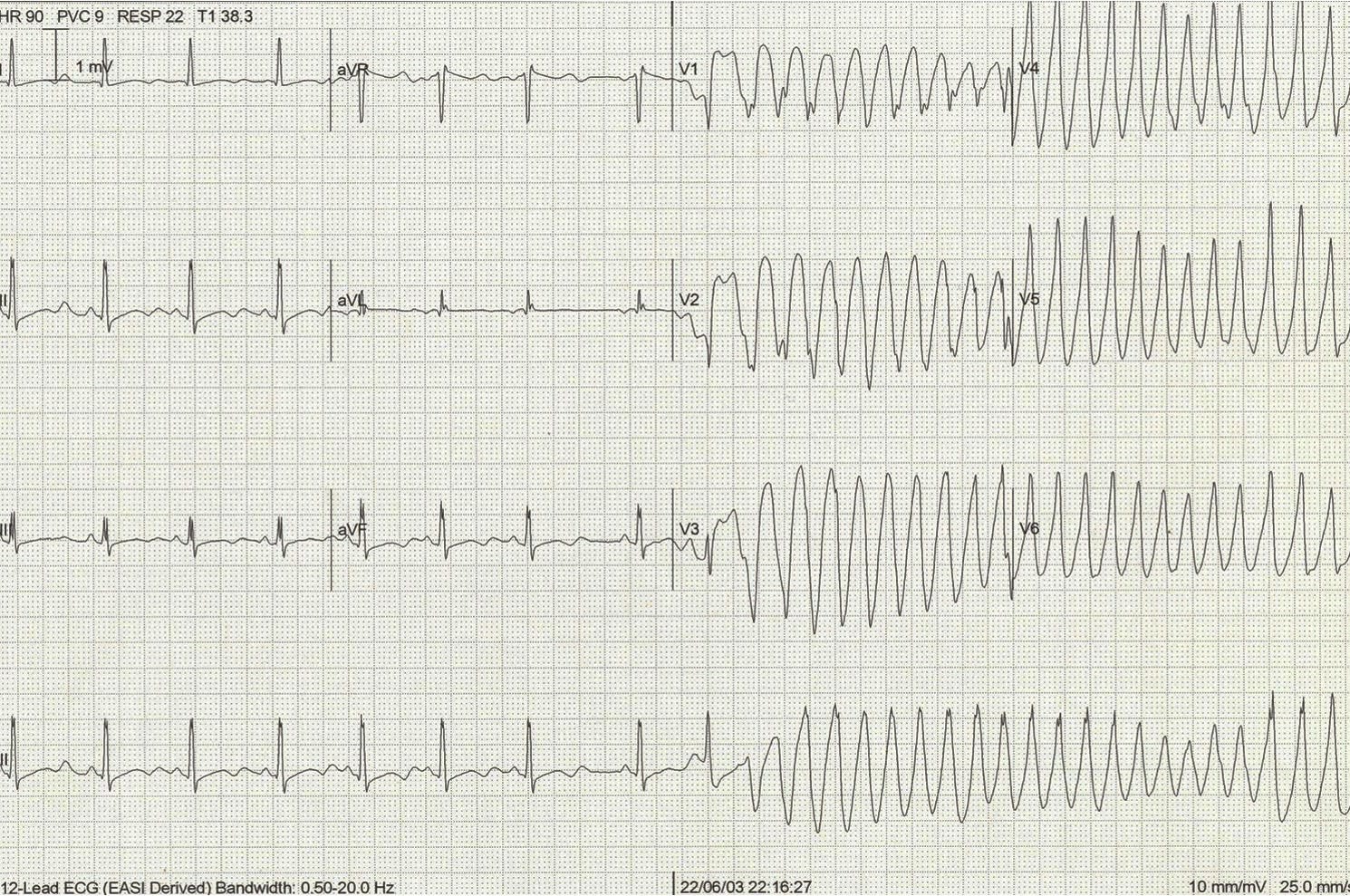
Prolonged QT going into monomorphic VT
Prolonged QR going into VF
Prolonged QT complicated but a severe movement artifact.
Prolonged QT going into polymorphic VT.
A prolonged QT interval has a long list of potential causes. In her case it was severe hypokalemia and hypomagnesemia. She went into Torasades de Pointes (Polymorphic VT) but surprisingly maintained a BP of 70systolic and spontaneously went back into sinus rhythm. Subsequently treated aggressively with K and Mg replacement.
A 26yr old Vietnamese man presented to ED following a sudden collapse at home. He is now well. What does the ECG show?
HOCM..
Anterior STEMI.
Brugada syndrome.
Sinus tachycardia and RBBB secondary to pulmonary embolus.
Brugada syndrome is a sodium channelopathy most commonly seen in people from South East Asia. They are prone to sudden VT and once proven should get an implantable defibrillator.
A 28yr old man presents with mild left sided pleuritic chest pain. The following ECG is performed. What should happen to the man?
Give aspirin and organise urgent cardiac catheterisation.
Thrombolyse ASAP.
Admit to CCU. Serial troponins and reassess tomorrow.
Repeat ECG and if serial troponins normal, discharge home.
The trick here is than the ECG is on the wrong voltage setting making the ST elevation more pronounced. He actually has benign early repolarisation and presented with pleurisy. (PS he was thrombolysed without incident as the ECG voltage abnormaility was not noted.)