Point of Care Ultrasound (POCUS).
A 20yr old man is involved in a high speed MVA and is hypotensive. What does the RUQ view show?
Free fluid in "Morrison's Pouch".
Free fluid in the Splenorenal angle.
Physiological fluid between liver and right kidney.
Clotted blood in between liver and right kidney.
There is free fluid seen as a hypoechoic strip on the ultrasound clip in Morrison's pouch. Morrison's pouch is the region between the liver and the right kidney.
This is an upper abdominal ultrasound clip and shows a "seagull" sign. What anatomical structure is this?
Superior mesenteric artery.
Inferior mesenteric artery.
Middle mesenteric artery.
Coeliac axis.
It is a pretty wonky looking seagull if you ask me. The coeliac axis (or trunk) comes off the aorta before the superior mesenteric artery.
What does this ultrasound of two Achilles tendons being dorsiflexed show?
Two normal Achilles tendons in people of different age.
The first Achilles tendon is normal the second shows degeneration.
The first Achilles tendon is normal, the second ruptured.
Both are ruptured, the second completely.
Note--in the first part of the video the normal tendon is highlighted with an orange bracket and can be seen to be sliding normally. The second part of the video has lower depth and the arrow points to the disruption in the tendon.
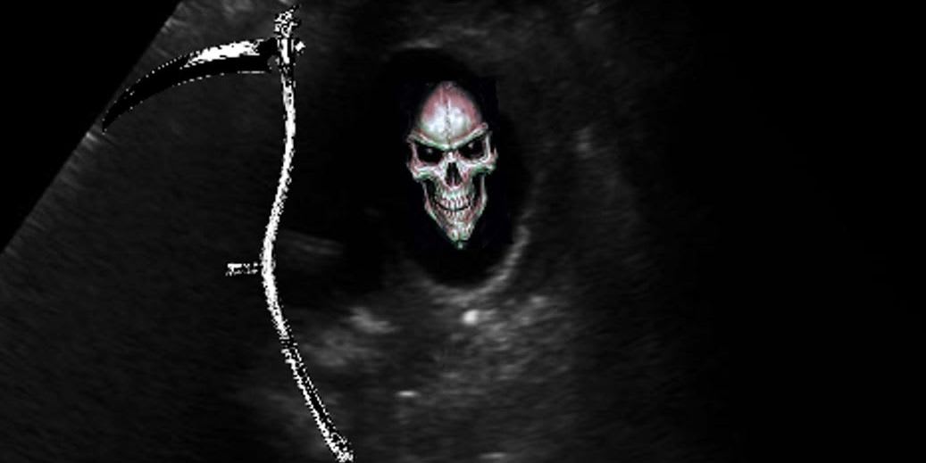
A 64yr old man presents with severe back pain. What does the ultrasound show?
A distended gallbladder with a valve.
An aortic aneurysm.
An aortic dissection.
An aortic dissection in an aneurysmal aorta.
The ultrasound clip is about at the level of the coeliac axis. You can see the aorta with a horizontal flap moving within it. You cannot see a thrombus and the aorta is not aneurysmal (ie it is < 3cm wide--measured outer wall to outer wall).
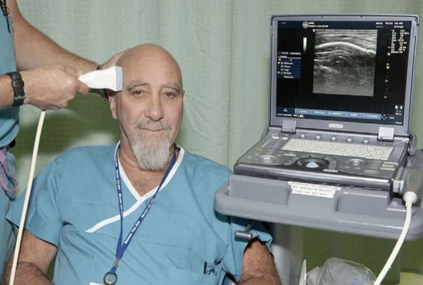
This man is taking part in a double blind trial in the University of Arizona looking at using trans cranial doppler to treat what condition?
Cerebral tumours.
Multiple sclerosis.
Chronic pain.
Perpetual smiling.
Dr Hameroff (pictured) is the lead author of a double blinded paper that used transcranial ultrasound on 31 patients and showed an improvement in chronic pain for up to 40min after 2mins of trans cranial ultrasound.
His interest was sparked when he did it on himself and noted a transient improvement in mood.
Go figure---but don't do it on yourself---someone will blame me.
His interest was sparked when he did it on himself and noted a transient improvement in mood.
Go figure---but don't do it on yourself---someone will blame me.
A 42yr old lady has right upper quadrant pain. Which answer best describes the findings?
Stone in the gallbladder neck. Murphy's positive.
Stones in the gallbladder neck, thickened GB wall with pericholestatic fluid.
Gallstone in the gallbladder. Distended gallbladder.
Gallstone in the neck of the gallbladder. Free fluid in Morrison's pouch.
You can see a few stones in the neck of the gallbladder with their post acoustic shadowing. The anterior wall is > 3mm and you can see an anechoic strip of fluid--the pericholestatic fluid. Ultrasound positive Murphys is when you poke the gallbladder with the probe and it is tender.
A 68yr old man present with chest tightness, diaphoresis and shortness of breath. The appearance seen in the clip is all over both lung fields. What does it show?
Widespead B lines consistent with pulmonary oedema.
Widespread B lines consistent with bilateral pneumonia.
Widespread A lines consistent with pulmonary oedema.
Normal base of lung.
B lines are artifactual and arise from the presence of fluid in the interlobular septae. (hydroaeric reverberation artifact). < 3 can be normal especially in the elderly and lung bases.
To be a B line it has to arise from the pleura, obliterate A lines. go all the way down the screen, move with the pleura and be well defined.
A lines are horizontal reverberation artifacts off the pleura.
To be a B line it has to arise from the pleura, obliterate A lines. go all the way down the screen, move with the pleura and be well defined.
A lines are horizontal reverberation artifacts off the pleura.
A 43yr old lady with a history of breast cancer presents with shortness of breath and hypotension. What does the video clip show?
A massive pulmonary embolism.
Biventricular failure.
A large pericardial effusion.
A large pericardial effusion with signs of tamponade.
You can see in diastole the right atrium completely collapsing. The right ventricle looks a bit like someone is jumping on the free wall---the trampoline sign. Both signs of cardiac tamponade.
This man has presented with shock of uncertain cause. What does this clip show?
A normal IVC with tachycardia.
An underfilled IVC with tachycardia.
An overfilled IVC with tachycardia.
An IVC consistent with a massive pulmonary embolus.
Normally the IVC is about < 2.1cm and collapses about 50% with inspiration. Here it is very thin and collapses almost completely with inspiration. This is consistent with hypovolemic shock.
A 24yr old man went bungy jumping then developed decreased vision in his eye. What does the ultrasound clip show?
A dislocated lens.
Ascaria lumbricoides.
Retinal detachment.
Vitreous haemorrhage.
That characteristic worm like dance on moving the eye is characteristic of a retinal detachment. You can see its attachment where it is tethered to the optic nerve.