Eyes.

This man developed a painful red eye on entering a dark room. He has photophobia and is nauseous. What does the patient suffer from?
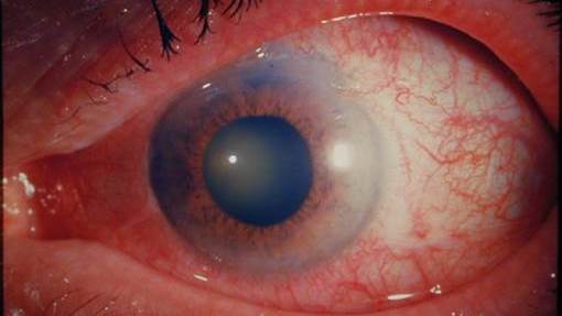
The pupil has dilated when the patient entered the dark room. This has thickened the iris which in turn has blocked the angle in the anterior chamber through which the aqueous fluid drains. Pressure quickly rises causing pain and the scleral injection seen. The pupil responds poorly to light and the cornea is cloudy.
This lady has suffered a blunt injury to the eye. What does it show?
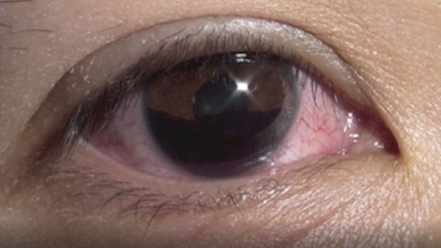
A hyphaema. The blood sits in the anterior chamber and is usually caused by bleeding from vessels in the ciliary body. Gravity often allows it to appear as a fluid level.
This lady has been asked to look up after a blunt injury to her left orbit. What muscle has been trapped making her unable to elevate her left eye?
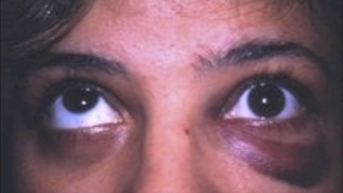
A fracture of the inferior aspect of the orbital floor can get the inferior rectus trapped. This stops the patient being able to gaze upwards on the affected side.
Arctic reindeers eyes change colour from gold in summer to blue in winter (pictured). Why is this?
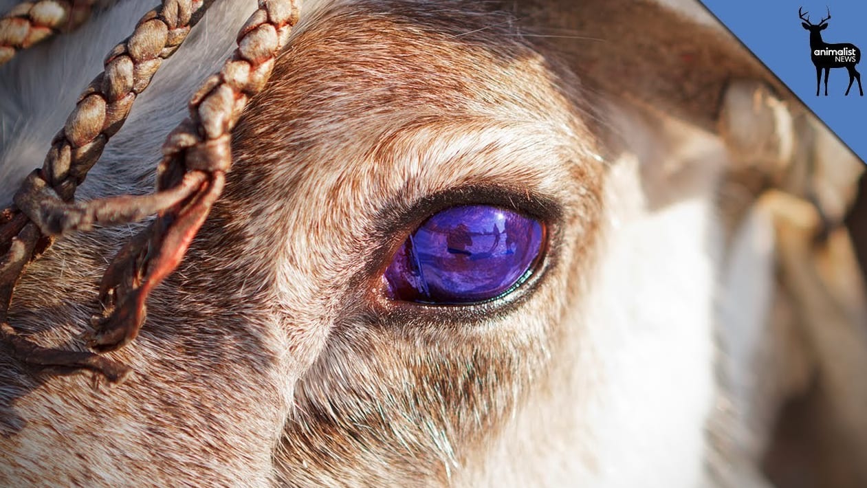
The part that changes colour is called the tapetum lucidum. It is a shiny, mirrored layer behind the retina that helps some animals to see in the dark.
When light enters the eye much of it hits the sensitive cells in the retina. But sometimes it misses the mark. The tapetum lucidum gives the eye a second chance to detect the light by reflecting it back towards the retina again.
In many animals this reflective layer shines gold, permanently. While reindeer eyes are also gold in the summer months, in winter their layer turns blue.
This lady has a warm tender left orbit and pain on eye movement. What is she suffering from?
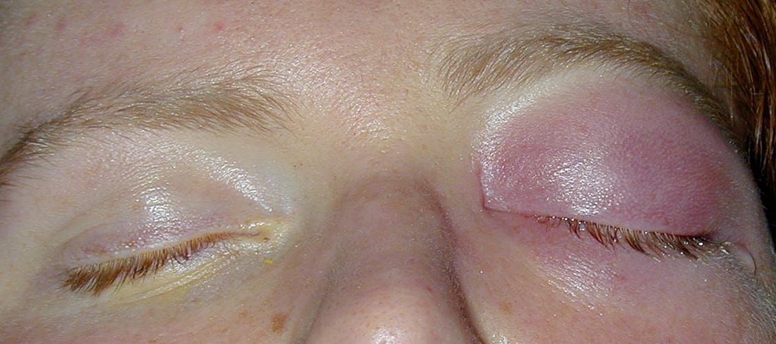
This is orbital cellulitis. The orbit has a fibrous septum which divides it into 2 parts---anterior to the septum called pre septal and posterior to the septum called orbital or post septal cellulitis. Orbital cellulitis causes proptosis of the eye as seen here.Can be a tad confusing.
This lady has Sarcoidosis and presents with a painful left eye along with photophobia. What does she have?
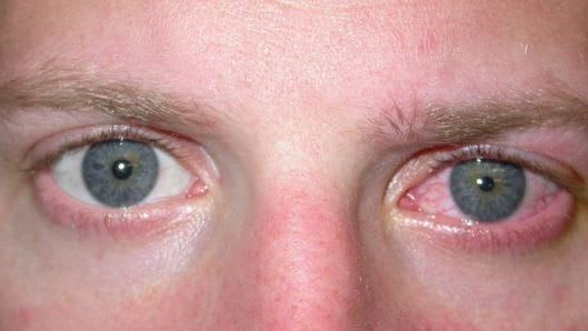
Iritis is also known as anterior uveitis. There are many different causes. Autoimmune disorders make up one group. Generally the treatment includes corticosteroid and cycloplegic drops (dilate the pupil).
This 69yr old man with a background of hypertension presented to ED with sudden loss of vision in one eye. This is what is seen on inspection of the retina. What has happened to him?
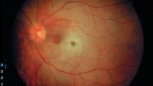
Sudden painless vision loss with a pale retina and prominent macula is a CRAO or Central Retinal Artery Occlusion.
This elderly man noticed a fibrous looking growth encroaching on his iris. What is it?
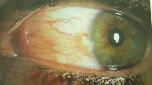
A Pterygium, which is "wing" in Greek, is a common benign growth of fibrous tissue that can be surgically removed. Note---Pangolins are the most trafficed animal in the world.
This middle aged lady presented with almost complete loss of vision in this eye. It is painless. What does the fundoscopy of the retina show?
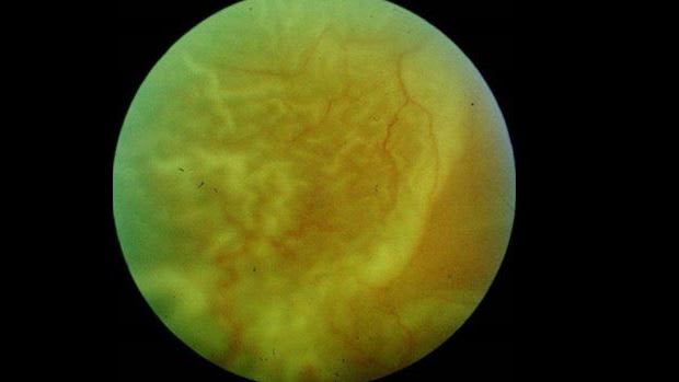
It shows the characteristic wringled appearance of an elevated retina. An ultrasound will easily show the retinal detachment.
The animal in the video is a Tarsier. What is true about its eyes?
Tarsiers can also rotate their heads 180 degrees in each direction and are the only entirely carnivorous mammal. Funky.