Click on the image you would like to win!
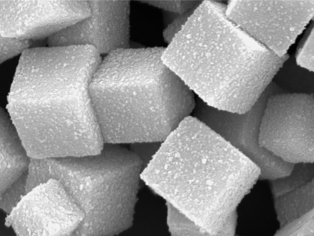
Iron Oxide coated with silver. Submitted by: Javeria Bashir
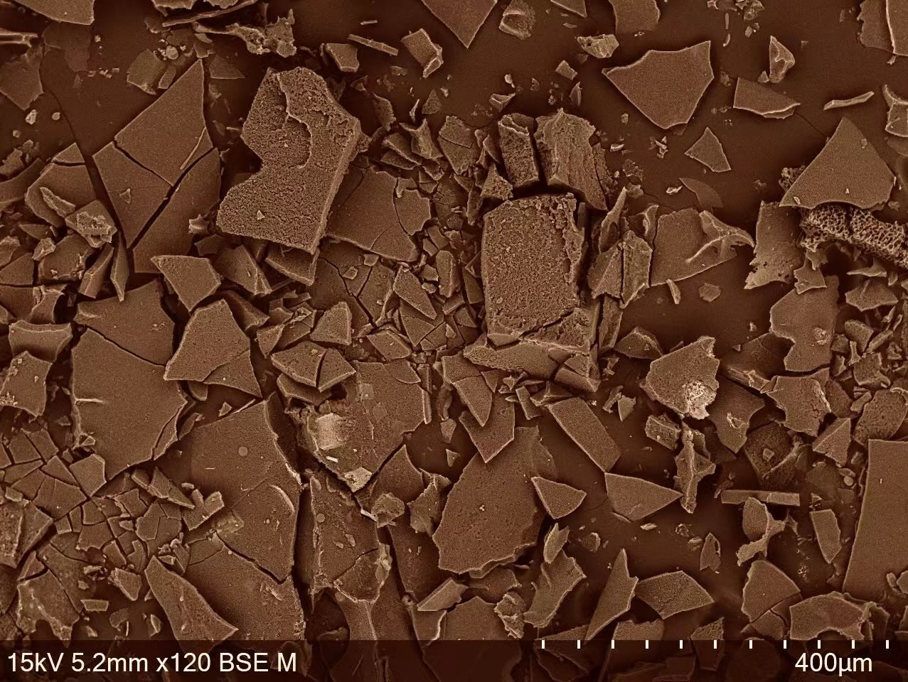
Microchocolate made from enzymes under a scanning electron microscope (SEM). Submitted by: Yilun Weng
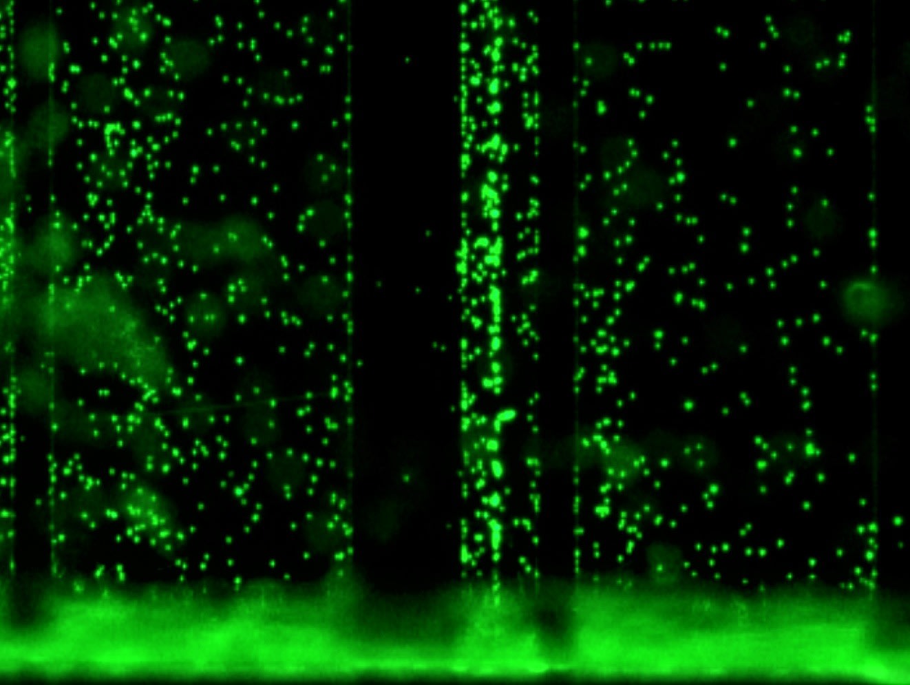
Not the Matrix, just the magnificent, fluorescent world of microfluidics. Submitted by: Raissa Teixeira
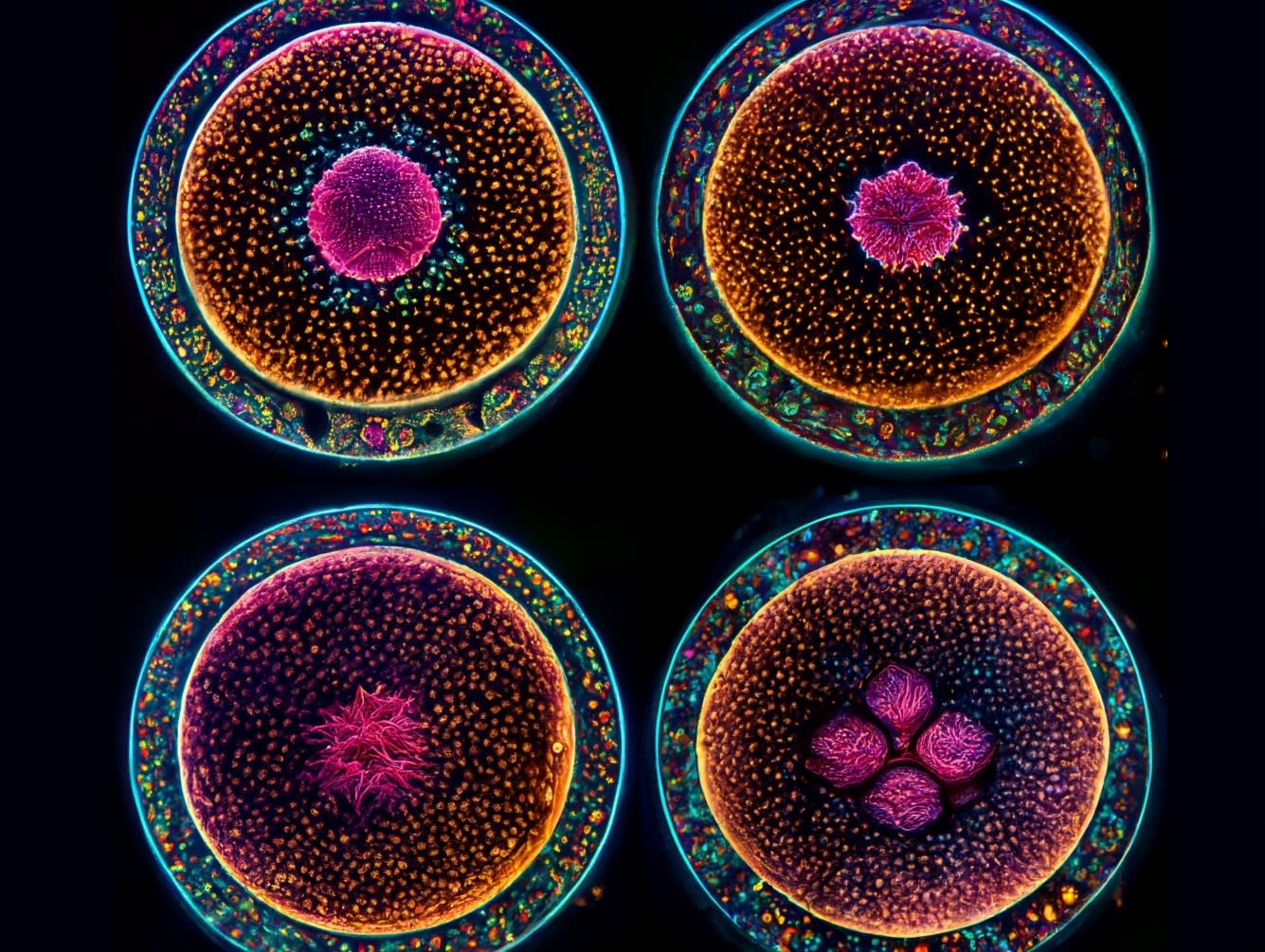
Computer aided design depiction of embryo development. Submitted by: Larry Cai
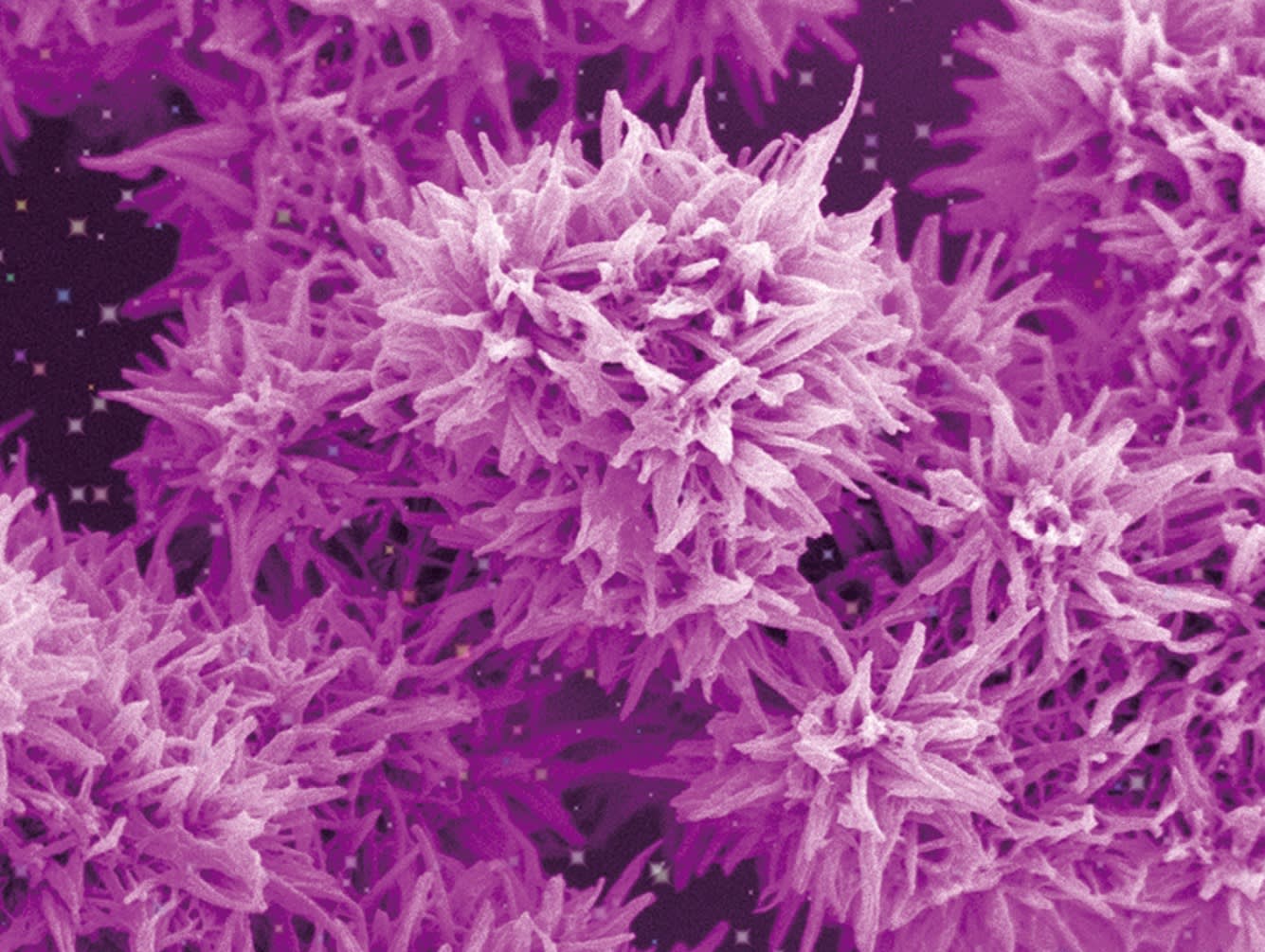
Flower-like titanium dioxide assembled by small rod-like particles. Submitted by: Yusuf Valentino Kaneti

Rough silcia nanoparticles taken at the Centre for Microscopy and Microanalysis housed in AIBN. Submitted by: Dan Cheng
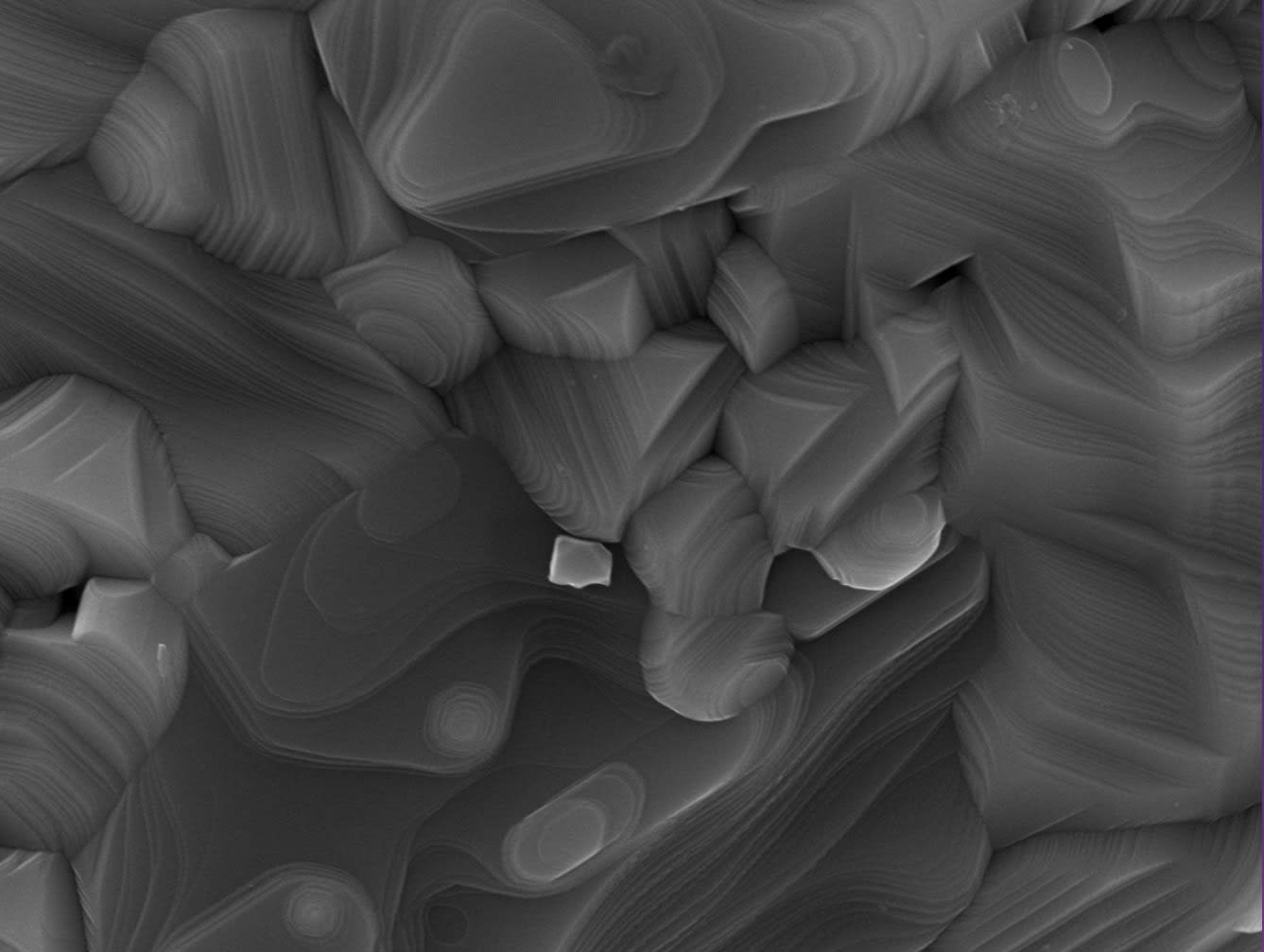
This image resembles a terraced field, which is a famous farming landscape in China.Here, we see it in nanoscale. Submitted by: Jiakang You
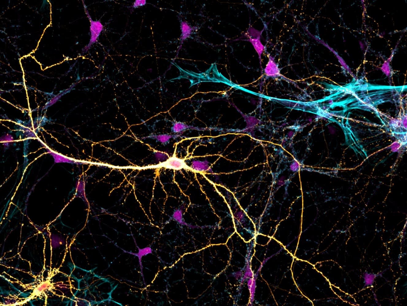
Hippocampal neurons stained with monomeric and polymerized actin and green fluorescence protein. Submitted by: Belal Shohayeb
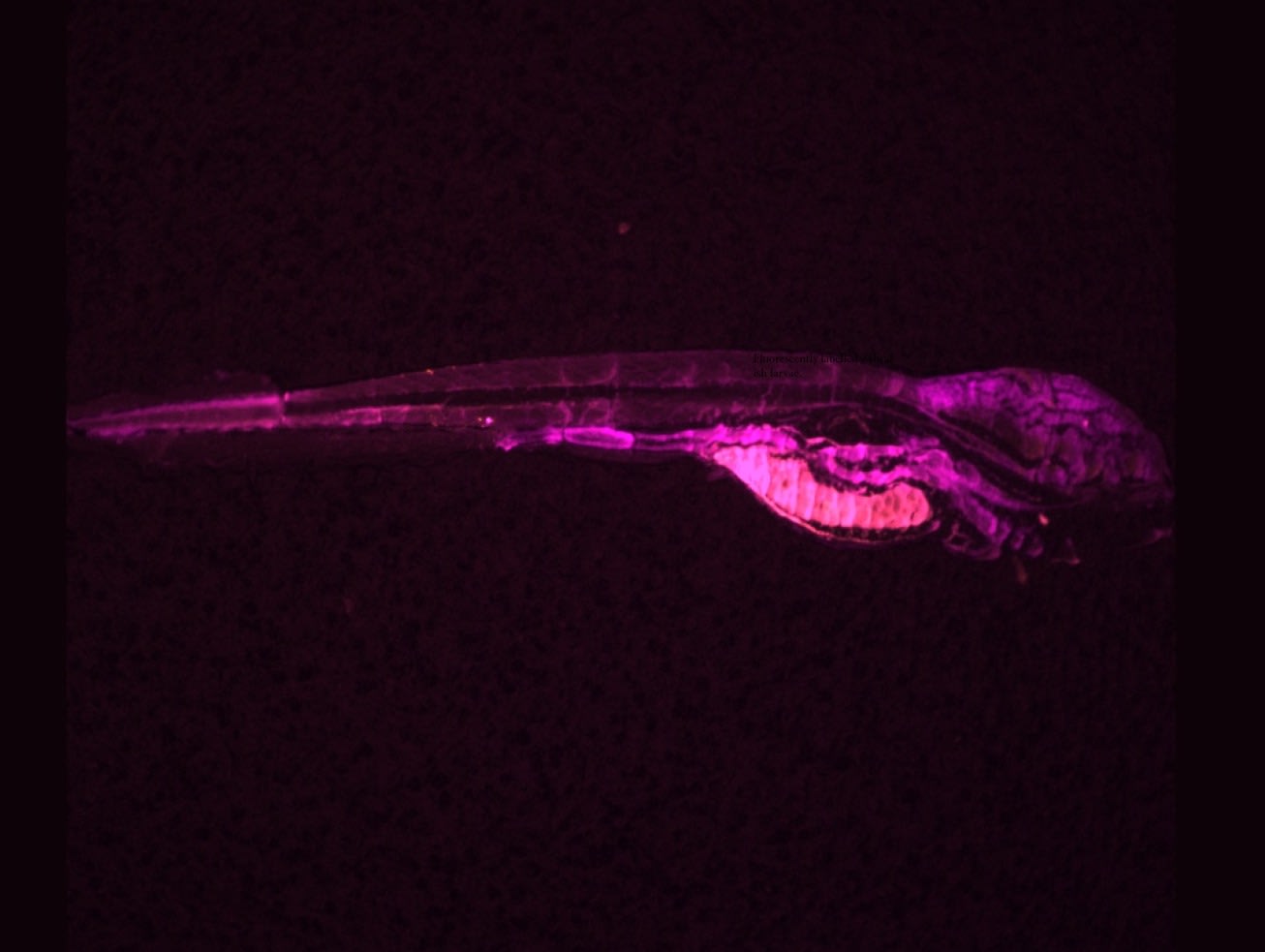
Fluorescently labelled zebrafish larvae. Submitted by: Syed Aoun Ali
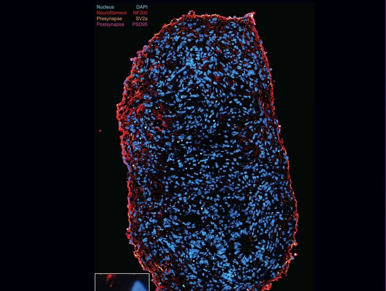
A 1-month-old spinal cord organoid showing early synapse formation between motor neurons. Scale bar 100 micron. ROI scale bar 2 micron. Submitted by: Sean Morrison
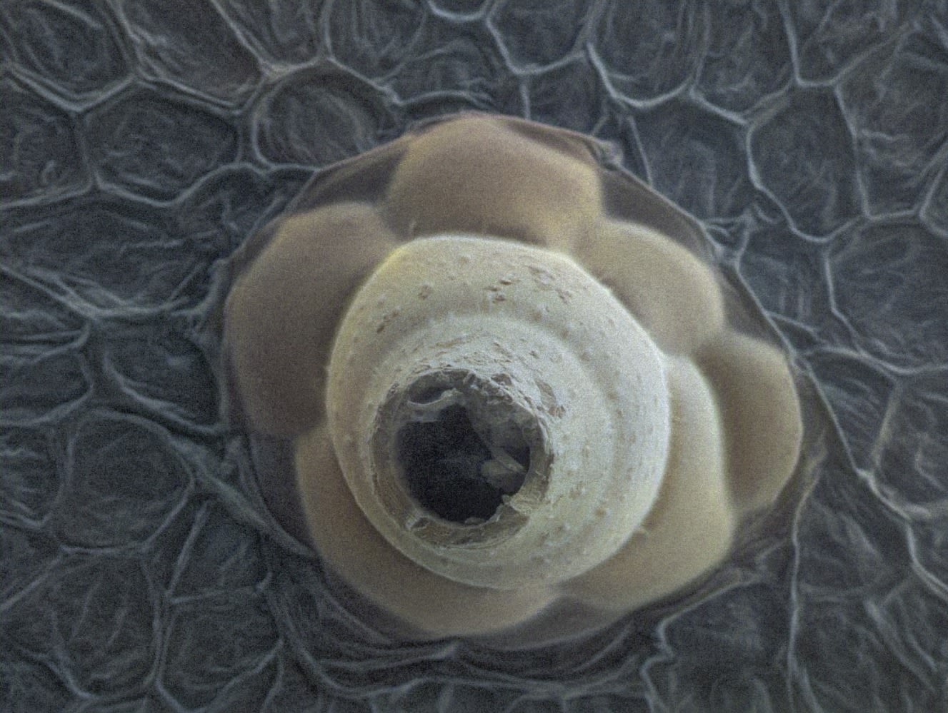
Scanning electron microscope (SEM) image of adaxial cucumber leaf. Submitted by: Shangxu Jiang
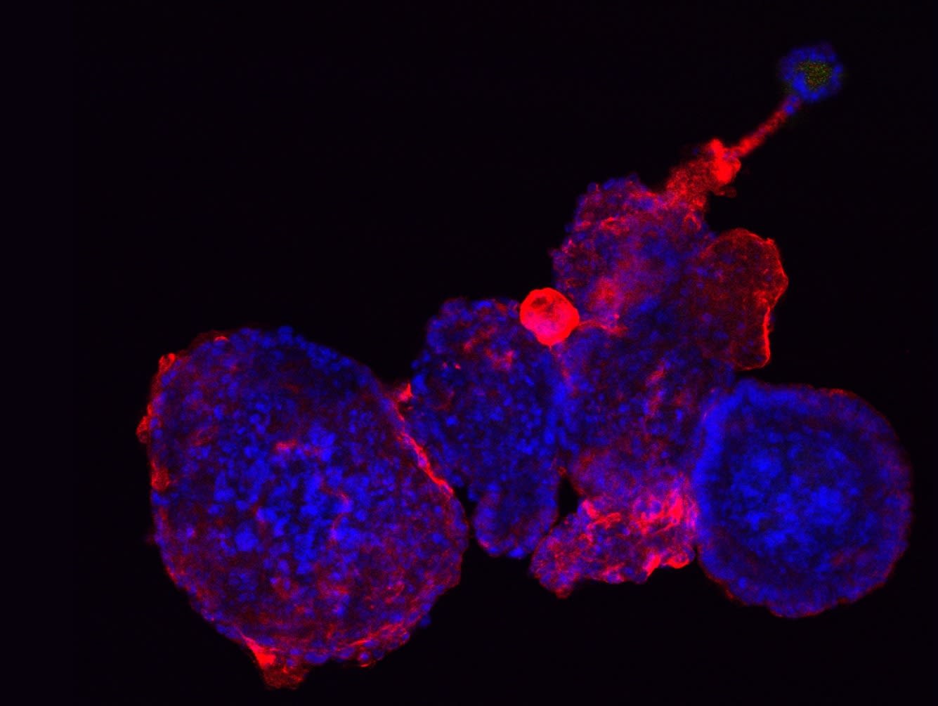
An intestinal organoid taken by confocal microscopy. This organoid differenciated into a motercycle! Submitted by: Jie Tang
Enter your email to sign up for our newsletter (optional)
Thank-you
Thank-you for voting!
Winners will be announced after the poll has closed via the AIBN social media pages